7: Mug ShotsOfficially they called it the Cell Culture Laboratory of the University of California at Berkeley, but in fact it was located in Oakland. Not in Oakland's fashionable residential hills but in the western flatlands, the marine industrial district. This was the part of town you saw first as you drove from San Francisco across the Bay Bridge: the storage tanks, the warehouses, the big Rustoleum red containerships at the docks, and, looming over it all, the gigantic metal cranes like a herd of mechanical dinosaurs foraging among the cargo at the water's edge. You knew you were in Oakland's harbor district when the roadside billboards stopped trying to sell you Johnny Walker Red and started pushing Tasco industrial valves.
The cell culture lab was housed in a former mess hall within a three-square-mile supply compound run by the U.S. Navy. There were no signs of Berkeley's rolling green lawns, the quads where students gather to picnic and play music, or the majestic marble buildings of higher learning. Here the office buildings were gray with dirty windows. Huge anchors and propellers were strewn around aluminum warehouses. In the alleyways stood dingy yellow forklifts. Scores of cargo containers, each the size of a Greyhound bus, were stacked high behind chainlink fences topped with barbed wire. Down the road was an outfit that hauled these "piggybacked" containers by rail and truck. It was called the Southern Pacific Golden Pig Service.
And yet inside the white building that held the cell culture lab there was no hint of the surrounding clutter. There was order and, through a certain set of double doors, there was cleanliness beyond imagination.
You couldn't go through the doors -- they were locked -- but you could peer through their windows into a bright, gleaming hallway. To get to that hallway you had to step into what looked like a large closet next to the double doors. Inside, a wooden bench ran from one side of the closet to the other, blocking your way. Where you entered, the floor looked like most institutional floors; its pattern of charcoal gray swirl was dulled by a hazy buildup. But just past the bench, it was a polished gem, the proverbial "floor you can eat off." On the wall was a large black-and-white sign:
Observe Change Rule
Never Come into Laboratory with Street Shoes Alone
The sign hung next to a series of shelves with forty-eight cubby holes, each labeled with the name of a lab worker, each holding a pair of shoes. Staffers had to change out of their street shoes and into a pair they wore only in the lab. For visitors there were white nylon booties in small, medium, and large.
The trick was to sit on the bench, put on a clean shoe (or slip a bootie over a dirty one) and swing it over to the clean side; then repeat the process for the other foot. Lab workers found the maneuver quite natural. But visitors often had trouble, accidentally bringing a clean bootie down onto the dirty side, in which case a new bootie was called for; or landing a dirty shoe on the clean side, which required a quick swabbing of the deck; or nearly falling off the bench in an attempt to avoid doing either.
The door in the clean section of the closet led into the gleaming corridor. Several individual lab stations extended along the hallway's left side. At the near end was the assay area, a large open space where newly arrived cells were logged in, examined without opening the flasks or dishes that held them, and stored until they could be analyzed more thoroughly and in the security of one of the "clean rooms" a little farther down the hall.
Each clean room was really a room within a room. The anteroom, about the size of three telephone booths, was where two technicians in surgical masks, caps, gowns, and gloves prepared for the task at hand, readying equipment, squirting alcohol over bottles of nutrient solution to kill bacteria, removing sterile wrapping on the glassware they planned to use. They then entered the pristine inner sanctum, closed the sliding door, and set to work: perhaps feeding a cell culture, transplanting portions of it into other flasks, or replacing the liquid medium with a fresh supply -- changing its diapers, as the crew described it. The inner room had its own source of filtered air that kept the pressure there higher than in the rest of the lab, preventing airborne undesirables from drifting inside. When the door was opened, the air always rushed out of the room, never in. The technicians worked in pairs because it eased the load of monotonous chores. Open the flask, withdraw 10 ccs of medium, close the flask, label a new flask, open it, dispense 10 ccs of medium .... Working in pairs they could also keep an eye on each other. They were not to talk unless absolutely necessary; in spite of masks, talking increased the chance of spitting out microorganisms that might endanger the cell cultures. To eliminate the risk of cross-contamination, they worked on one cell line at a time, re-sterilizing the whole show before another line was brought in. It was an expensive, time-consuming procedure. They stayed in there for two to three hours at a time. But then obsessive care was what made Oakland so special.
When work on a particular culture was finished, any materials to be discarded were placed in pans full of disinfectant and taken to the far end of the hallway to the autoclave, a little chamber of hell that subjected its contents to lethal temperatures and pressures. All glassware was sent to be washed and rinsed in scalding, deionized water. Every working surface in the clean room was then wiped down with alcohol. Just in case some scrap of life remained at large in the room, and to kill off anything that might creep in later on, one of the technicians flipped a switch on the way out, bathing both compartments in ultraviolet light. On the door to each clean room was a sign that said in red letters:
KEEP CLOSED
Admittance to authorized personnel only.
Visitors and personnel not assigned to this area
contact W. Nelson-Rees.
On the other side of the corridor were the cramped quarters where Nelson-Rees and his co-workers mulled over the results of the lab work. Bob Flandermeyer had the office next to Nelson-Rees. He did most of his mulling while hunched over a table, pawing through a pile of photographs like a man searching for the right piece in a jigsaw puzzle.
***
Chromosomes, chromosomes, you could always find Bob Flandermeyer at that table sorting through hundreds of black-and-white enlargements of chromosomes. Long, straight chromosomes; short, stubby chromosomes; chromosomes that looked like bow ties, black ants, and licorice twists; and all wearing stripes, the irregular barber pole stripes known as banding patterns. Slowly, deliberately, ploddingly, Bob Flandermeyer would cast his big soft eyes over each photograph, studying the shapes and patterns.
Flandermeyer was a bear of a man with blond hair and a wide, friendly face. He spoke softly and a little ponderously. His laugh, loud and sharp, was startling punctuation to his sleepy speech. In early 1973, he left his job as a technician in a neighboring Navy laboratory. He had been working on a study of how bacterial diseases spread among troops, which had little to do with cell biology. Nevertheless, Nelson-Rees hired him to help apply the technique of chromosome banding to the identification of cells. Two other lab assistants before Flandermeyer had tried and failed to make it a practical system. The work was too tedious for his predecessors, but Flandermeyer mastered it.
The first part of the technique was nothing more than what karyologists, chromosome analysts, had been doing for years. The idea is to catch the cells in a stage of their growth cycle called metaphase, when the chromosomes line up across the center of each cell in orderly rows. You do that with colchicine, a drug derived from crocuses. Colchicine prevents the formation of tiny fibers that normally attach to the chromosomes and drag them to opposite ends as a cell prepares to split. It halts the chromosomes in their lineup like dancers in midstep. Then you add a solution to make the cells swell, which spreads the chromosomes apart so they don't overlap, and then an alcohol "fixative" to kill the cells. The next step is to attach a drop full of cells onto a microscope slide and flatten it into a layer thin enough that everything will be in focus for the camera. One popular method of flattening was the five-foot drop: holding the dropper at eyelevel, you aim at a group of slides arranged around your feet. From that height, a drop spreads itself into a broad, thin film, and if you're lucky the film ends up covering a slide instead of your shoes or the linoleum. For those with more flamboyant tastes, there was the crepe suzette technique: a match is put to the slide, igniting the alcohol fixative, which burns away in a few seconds, dramatically reducing the volume of liquid in the drop. Once the sample is properly flattened, you mix in a little Giemsa stain and place the slide under a microscope. The chromosomes show up in silhouette, dark blobs without detail. Based on their shapes, a good karyologist can tell the species of the donor: man, mouse, or whatever. By studying the shapes and counting the number of chromosomes, he can also tell if the cells are abnormal. Victims of Down's Syndrome, for instance, have one more than the usual human complement of forty-six chromosomes.
That was conventional karyology.
In the new banding technique, before staining the cells, you dip a slide full of them into trypsin, an enzyme that eats away parts of the protein coat over the chromosomes. Then, instead of painting the entire outer coating of the chromosome, the Giemsa stain is absorbed into it, but only in certain spots, creating discrete bands of darkness next to blank areas. The problem is that the trypsin and the stain have to be at particular concentrations and temperatures, and the slides have to be dunked into each solution for precise periods of time. Even if you figure out the proper combination, the technique doesn't work unless you catch the cells at just the right moment in the metaphase stage. For some reason, the enzyme can't eat through the chromosome's coating otherwise.
Flandermeyer studied several days with a group of researchers who were using the technique at the University of Indiana. He returned to Oakland and began experimenting. Time and again he would prepare a bunch of slides, dip them in trypsin while counting off the seconds, then dunk them in the stain, only to find the familiar dark blobs where he had hoped to see barber poles. That meant he had exposed the cells to the enzyme for too long, allowing the stain to seep in everywhere. Or had he got the temperature wrong? Other times, when he didn't keep them in long enough or failed to catch the cells in metaphase, no readable patterns appeared.
Finally he hit on a reliable recipe for producing good, clear bands. He stained stacks and stacks of slides, then sat down at the microscope and scanned one after-another, searching for the most photogenic cells. He photographed hundreds of them, printed up enlargements, cut out each banded chromosome, and mounted them all on white cardboard, grouped according to their banding patterns. After that the real work began.
First he had to memorize the patterns of normal human chromosomes. Every healthy cell has two copies of chromosome numbers 1 through 22, one from each parent, plus either a pair of Xs in the case of females or an X and a Y for males. So there are twenty-four different types of chromosomes, each type absorbs the stain in a characteristic set of bands -- its mug shot -- and Flandermeyer had to know each one on sight. He stared at the photographs for weeks. Through brute force of will he developed an eye for all twenty-four. But because these were normal chromosomes, present in every human cell, they were really no help in telling cell lines apart -- not directly. He needed to know the normal chromosomes only so that he could recognize the misfits, the ones with funny shapes and weird banding patterns.
The misfits, the theory went, resulted from random errors in the duplicating process. Such errors occur frequently in tumor cells, where the usual mechanisms of replication and growth run amok. The lower arm of a number 5 chromosome might break off and attach itself to a severed upper arm of a number 3, for example. This unlikely goat with a lion's head, passed on to daughter cells, becomes a unique marker of that particular tumor and of any cell line derived from it. The odds were slim that an identical misfit would arise by chance in another cell line. And it was close to impossible for one cell line to randomly create a set of three or four misfits that matched those in another culture. Only cells with a common origin would have the same aberrant marker chromosomes.
For Nelson-Rees, who had always been partial to chromosomes, and who had learned the value of misfits from Mr. Chromosome himself, these markers were among the more powerful means of identifying human cell lines. When the National Cancer Institute made the Oakland lab its hot new repository for human cultures, he decided that his quality control arsenal had to include a method of recognizing markers. But conventional staining wouldn't do it. To spot a marker you have to be able to make out which pieces of normal chromosomes had combined to form it. For that level of detail, you need to see the bands. That was why Nelson-Rees needed Bob Flandermeyer. And that was why Bob Flandermeyer, having learned every normal banding pattern, began staring at photographs of the abnormal markers, forcing his mind to associate each strange patchwork pattern with the cell line it had come from. The first markers he learned to spot were the four that other researchers had found in HeLa cells.
My God, he's slow, thought Nelson-Rees as he watched Flandermeyer pick his way through the photographs of unidentified cell cultures, looking for markers he had already memorized and memorizing the ones he came upon for the first time. "Ein Stier," Nelson-Rees had once called him, "an ox."
There was considerable art to the method, though. Flandermeyer knew that a banded chromosome's appearance often depended upon the angle from which he photographed it and whether it happened to be drooping or twisted or contorted in some other way. There was a lot of visual judgment involved, and Flandermeyer was not one to jump to conclusions.
Nelson-Rees and some of the other researchers who had learned to read banded karyotypes with Flandermeyer's help would often ask him to confirm their own assessments. He was, after all, the expert. On these occasions, Flandermeyer would stare at the photographs endlessly as the rest of them paced about. Finally, when he had convinced himself that yes, here was a marker chromosome in a suspect cell line, a marker that he recognized -- but only when he was dead sure -- he would smile broadly and say, "Well, lookie here."
With those words, Flandermeyer had helped Nelson-Rees finger the Russian cell cultures as HeLa contaminants in the fall of 1973. Like a dogged assistant detective, he had pored over mug shots of the Russian chromosomes for hours until -- "Well, lookie here" -- he found all four of HeLa's known marker chromosomes. The Russian case had been their first practical application of the banding technique. Now, a few months later, Flandermeyer sat in his office, methodically sorting through the mug shots of two more cell lines of questionable identity. One was a line of breast cancer cells called HBT3, HBT for human breast tumor; the other was a culture of human embryonic kidney cells designated HEK.
Nelson-Rees had become suspicious of the two cultures several weeks earlier after walking in on a conversation between Adeline Hackett and Trudy Buehring, two researchers at the cell culture lab who worked independently of his cell bank. Hackett and Buehring were talking about this puzzling culture they had come across while studying the common structures of breast cancer cells. Spread out on the counter in front of them were photographs of cells from a number of established breast cultures. The pictures, taken through an electron microscope, showed that one of the cell lines, HBT3, had none of the features found in the three or four others.
"They just don't look like breast cells," Buehring was saying. "They don't look like normal breast cells, and they don't look like cancerous breast cells."
"Isn't that strange?" said Nelson-Rees. Then he remembered that he too had seen something funny in HBT3, almost a year earlier, when a California researcher under contract to the institute sent him a sample for analysis and deposit in the bank. Flandermeyer had not yet joined the staff and the only means of checking chromosomes was the conventional staining. Nelson-Rees's examination confirmed that HBT3 cells were of human origin, and that was as far as it had gone.
Except that he had seen an oddly shaped chromosome. His conventional staining had shown no detail, but the outline of the thing clearly resembled two little ears. Someone had called them Mickey Mouse ears. And come to think of it, he had seen a somewhat similar set of mouse ears in another cell line three years before that, in 1970. They were in a line of embryonic kidney cells called HEK. Maybe this oddball cell of Hackett and Buehring's doesn't look anything like a breast cell because it is actually from a kidney, thought Nelson-Rees.
He hurried off to pull the old sample of HEK out of the deep freeze and get Flandermeyer working on a comparison of the two cultures. The banding technique would show for certain whether the ears in one were the same as the ears in the other.
***
"Well," said Flandermeyer after an eternity, "lookie here."
The banding patterns of the mouse-eared chromosome in HBT3 precisely matched the patterns of the one in HEK. But that wasn't the end of it. There was another abnormal chromosome in HBT3, a new marker that Flandermeyer had never seen before, a long and boldly striped thing whose twin brother was smiling out at him from HEK as well. Two markers in common made an even stronger case that these were the same cell line. But ... what's this? Something else? Uh-oh, lookie here: four more weirdos that Flandermeyer recognized instantly.
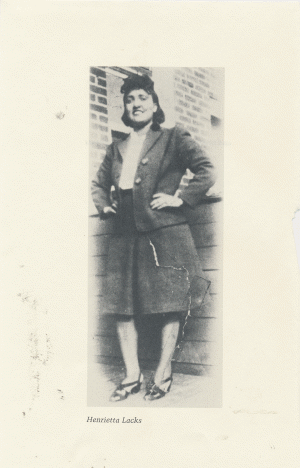
Nelson-Rees was amazed. Buehring and Hackett were flabbergasted. It appeared that HBT3 and HEK had been mixed up, as Nelson-Rees had suspected, but neither cell line retained its original identity. Both cultures were now HeLa, actually a previously unknown substrain with two new markers, but HeLa just the same. Instead of a cancerous breast culture and a normal kidney culture, what they had stumbled onto -- and what many investigators interested in breast cancer and kidneys were no doubt wasting their time studying -- were those familiar cervical cancer cells of Henrietta Lacks.
They were still reeling two weeks later when Flandermeyer emerged from a long session of mulling over mug shots to deliver more shocking news. Cell line HBT39B, yet another culture of supposed breast cancer cells that had been sent in for a routine check, also displayed the same six marker chromosomes.
It was like that point in every horror movie when the characters know there's no escape. True, there were no Saint Bernard-sized lumps of HeLa cells blocking the exits and gnawing through the telephone wires, but this horror story was for real.
First the Russian cell lines and now this. Three cultures from American scientists, cultures effectively selected at random, all taken over by the runaway cells of "our lady friend," as Nelson-Rees had started calling Henrietta Lacks. It reminded him of a remark made a few years earlier by a group of Johns Hopkins researchers who were marveling at HeLa's tenacity. "HeLa," they had written, "if allowed to grow uninhibited under optimal cultural conditions, would have taken over the world by this time."
The director of some other cell bank might have thrown out every culture of HBT3, HBT39B, and HEK, and left it at that. But to Nelson-Rees it didn't make any sense to merely note the problems, keep the cell bank pure, and let the calamities unfold elsewhere. Being the perfectionist that he was, Nelson-Rees couldn't stand the thought of all that error and confusion, not to mention the time and money undoubtedly being squandered by HeLa's victims. Being the keeper of the cells for the institute, the man armed with the latest techniques of cell identification, he felt almost duty bound to sound the alarm, to track down the fugitive cells of Henrietta Lacks, and to set things straight.
This was the quiet beginning of the crusade. There was no formal declaration. Nelson-Rees never called the troops together to say, "The war is on," and map out strategy. What he did, quite simply, was throw the entire operation into high gear. He began by tracing the path of the first breast culture. HBT3 had come to Oakland from a researcher at the California State Health Department, who, Nelson-Rees discovered, got it from a scientist at the Centers for Disease Control in Atlanta, who got it from Robert Bassin, an institute scientist, who originated the cell line in his Bethesda laboratory. Nelson-Rees wrote to all three. "Fully realizing the embarrassment to the originators of these cell lines and/or to the investigators from whom I obtained them, I would be very pleased to discuss this matter in detail with you to get at the source of this contamination, if indeed this is what is shown," he wrote. "I welcome and in fact must insist on further analysis of these and 'related' cell lines . . . ." It was like a note from the school nurse informing the parents that little Darlene had VD, and it drew the kind of reaction you'd expect.
Bassin was the first to respond. Being a careful scientist, he was well aware of the threat of HeLa contamination, which is why he had kept HeLa and all other human tumor cells out of his laboratory when he established HBT3 in 1972. Furthermore, his lab was housed in building 41, the institute's Emergency Virus Isolation Facility.
Building 41 was cut off from the world. No agent from the outside environment could leak in to jeopardize the purity of the experimental conditions there; none of the viruses or other nasty things they handled inside was able to leak out. There were no windows in building 41, no doors that opened without special clearance. The flow of air throughout the building was carefully controlled. According to institute legend, the high security so impressed a young medical intern who worked there one summer that it later moved him to write a science fiction novel about a deadly germ from outer space, The Andromeda Strain. In fact the book's author had never set foot inside building 41, but the facility's hermetically sealed atmosphere inspired those kinds of stories. It also made Bassin confident that Walter Nelson-Rees didn't know what he was talking about. Bassin telephoned to tell him so.
First of all, Bassin argued politely, it is very difficult to prove scientifically that two things are the same. It's simple to say they are different, of course. All you have to do is find some characteristics they don't have in common. But the fact that a few funny-looking chromosomes appear in both HBT3 and HeLa doesn't prove they are the same cell. Bassin questioned Nelson-Rees about this method of examining banded chromosomes. Could he be sure the markers matched identically?
Quite sure, said Nelson-Rees, adding that HBT3 was also carrying the A type of the G6PD enzyme, the type carried by HeLa cells.
That didn't prove a thing, Bassin came back, since the woman whose tumor established the HBT3 line was of northern Mediterranean extraction -- Greek or Italian. Although it happened very rarely, type A had been known to show up in these populations. And even if the culture of HBT3 in Oakland truly was HeLa, wasn't it perfectly possible that the ones he worked with in Bethesda were bona fide? Maybe the California scientist who sent the cells in to be checked had contaminated them with HeLa in his own lab. Or maybe it happened in Atlanta.
Perfectly possible, Nelson-Rees agreed, which was why he needed to examine a culture from Bassin's personal supply.
The following day, a shipment of breast cancer cells took United Airlines flight 57 from Washington, D.c., to San Francisco. A messenger delivered them to Nelson- Rees, who passed them on to Flandermeyer for analysis. Nelson-Rees also shipped a sample to Ward Peterson in Detroit for the G6PD testing. As he waited for the results, he continued writing letters, making phone calls, notifying and debating researchers who were connected with the three contaminated cell lines. Like Bassin, none of these researchers had been working with HeLa, or so they claimed. None put much credence in the method of chromosome banding. And those who conceded there might be a problem unanimously pointed the finger elsewhere.
Ernest Plata, the man who had originated HBT39B, the other breast culture, had considerably more in common with Bassin than any of the others. He had started his cell line a few months after Bassin had got HBT3 growing. And he had done it just down the hall from Bassin's lab, in the institute's windowless fortress.
Well, well, what a coincidence, thought Nelson- Rees. It was obvious to him that a HeLa culture was running around building 41, masquerading as at least one other cell line. And while the HeLa cells couldn't leak out the carefully monitored vents or the air-locked doors, they were far from trapped. Every so often Bassin, Plata, or perhaps some other unwitting accomplice of Henrietta Lacks would wrap up a few samples of contaminated cells and mail them off. Nelson-Rees would have to have the test results to be sure, of course, but it looked as though the National Cancer Institute was distributing HeLa cells, under various false names, all around the country.
The last member of the triad, HEK, Nelson-Rees could not trace back to its source. It had been established ten years earlier by a laboratory that no longer existed. Although it had been widely used in research, there were no records of the donor's race, sex, or any other characteristics that could have been checked by his chromosomal and biochemical techniques. The earliest cultures he could find were in the hands of researchers at Pfizer Laboratories in Maywood, New Jersey, who had had them since December 1964. He asked for a sample.
By February 1974, two months after Nelson-Rees and colleagues had found the first indications of this three-way contamination, they had called in and analyzed two or three specimens of each cell line. In every one of them, Peterson had detected type A G6PD. In everyone of them, including Bassin's personal supply of HBT3, Flandermeyer had found the marker chromosomes -- the four traditional markers as well as Mickey Mouse and the one with the bold stripes, which they had named the Zebra.
Because this strain of HeLa had developed two markers not present in other known HeLa cells, Nelson-Rees and Flandermeyer decided that it must have been evolving on its own for some time, perhaps in an isolated environment such as an institute laboratory. Personally, Nelson-Rees suspected that this variant of HeLa had first contaminated HEK, the old-timer, and then gone on disguised as HEK to spoil HBT3 and HBT39B. But such reconstructions were a secondary concern at the moment.
It was time to get the word out. In the few months it had taken to track these three lines, they had come across two other popular cultures that were contaminated with HeLa -- one a line of prostate cells, the other a culture of liposarcoma, a tumor of fatty tissue. There seemed to be HeLa contaminants everywhere they turned, though few people aside from the researchers Nelson-Rees had contacted had any hint of trouble. Nelson-Rees was still pushing, unsuccessfully, to publish the very first HeLa mix-up they had uncovered, the case of the Russian cells. Originally he had wanted to use the Russians' misfortune as a warning that HeLa might yet be alive and lurking around American laboratories as well. That warning was now well behind the times. These latest findings about five American cultures demanded some kind of all-out emergency alert.
"Anybody interested in working with characterized cell lines of bona fide purity of origin would be interested in this article," he wrote to Philip Abelson, editor of the journal Science. "Knowing that cell cultures presumably derived from human embryonic kidney, human breast carcinoma, human prostate tissue, and human liposarcoma cells are indeed derived from a human cervical carcinoma would certainly change the course of a number of research projects now in progress, and alter the interpretations in many publications already in existence involving these cells."
Science sent his manuscript to their technical reviewers, one of whom criticized Nelson-Rees's writing style, though he said he was sure that the findings were correct. The other said, "The main message of this paper is extremely important: that a surprisingly high proportion of cell lines are not what they are purported to be." In view of the rapidly increasing use of cell lines in research, the reviewer added, it is vital that Nelson-Rees's message be widely disseminated. From those comments, the editors at Science somehow decided that the report didn't quite meet publication standards. No thank you, they wrote back to Nelson-Rees.
How's that? Not interested in publishing the news that five cell cultures widely used in cancer research today are not what they're supposed to be? Was it happening again, just as it had with the Russian cells -- this dead silence, this dumb stare, this gaping lack of interest in what he knew to be findings too incredible to ignore? Not this time. No, this time the sheer shocking momentum of his results couldn't be stopped. Rumors were already circulating. At the urging of several scientists and friends who had heard them, Nelson-Rees re-submitted the manuscript to Science.
It was then that Bob Bassin did a brave and unusual thing. After studying Nelson-Rees's banding data, and having performed a few tests of his own, Bassin conceded that his HBT3 cells might well be HeLa. This was no private confession. Bassin wrote to twenty researchers around the world to whom he had sent samples of his cell line, informing them of the bad news and asking that they send copies of his letter to anyone they had shared the cells with. Among other things, it was a graphic illustration of how far such an error might perpetuate itself.
Bassin also sent a copy of the letter to Nelson-Rees, who forwarded it to the people at Science with a note saying, "You will, no doubt, appreciate the need for our publication." This time they appreciated it. The manuscript was approved and rushed into type. Nelson-Rees dictated a revised introduction and checked the galleys by telephone.
A few weeks later he was in Miami, attending the annual meeting of the Tissue Culture Association, when Science mailed out the issue carrying his report. A desperate fellow approached him at the pool of the Hotel Deauville, where the conference was being held. The man looked deeply troubled, as if he had just learned that tomorrow after breakfast the universe would blink out of existence.
"As I left the lab today," the man said blankly, "I saw the Science article. I just couldn't believe what I read."
Nelson-Rees had no idea how to respond. The lost soul turned and wandered off behind the lounge chairs and umbrellas.
Suddenly people were stopping him in the hallways to ask about chromosome banding and dropping by the dinner table to check a point about sterile procedures. The next day the place was positively buzzing about HeLa contamination, and the conference organizers asked Nelson-Rees to deliver an impromptu talk on his work. He didn't hesitate.
At last, he had got someone's attention.